
Fig. 2.
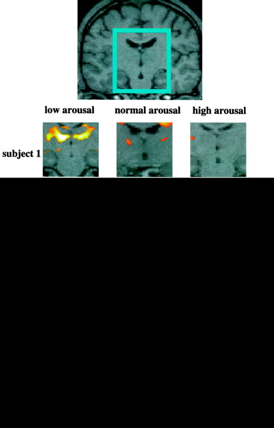
Activation in the left and right thalamic regions (centered in the inset) in each subject in the contrast attentional task versus passive viewing in normal, high, and low arousal. Note the greater activation when the test is performed in a state of low arousal. In each subject the areas of activation are superimposed on the correspondent structural images (which are co-registered to the functional ones). This procedure permits greater sensitivity in identifying anatomical structures because there is no need to account for variations in normal anatomy between subjects. The apparent activations in two subjects in the ventricles are probably artifactual. The Z-value represents the degree of significance of the activations (see Materials and Methods).