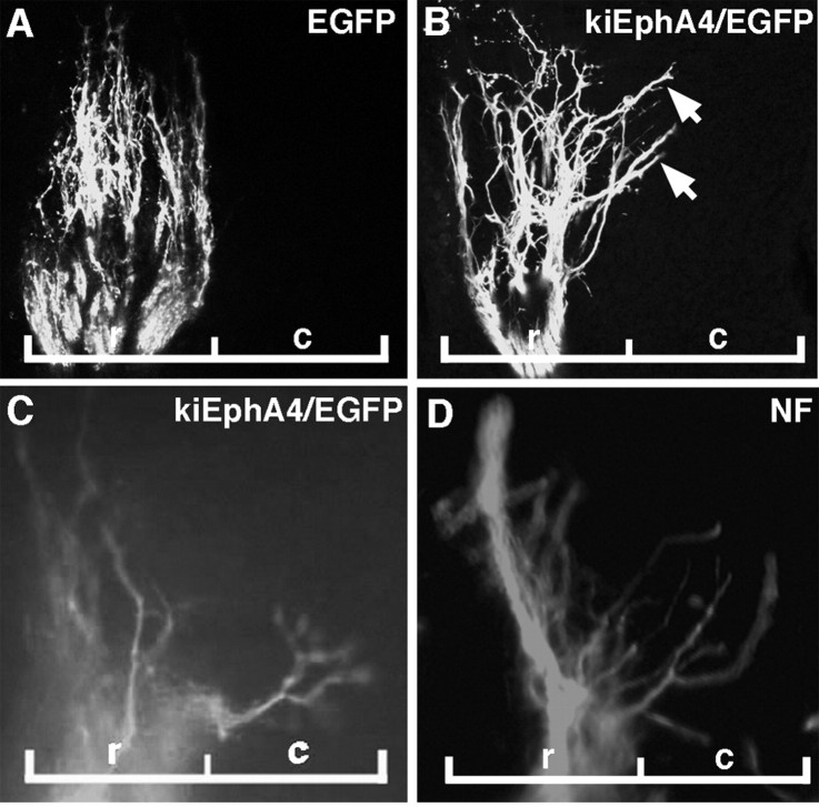
Figure 4.
MMC(m) axons grow aberrantly into the caudal half-sclerotome when EphA4 signaling is blocked. All images are sagittal sections through MMC(m) axons, at the level of a single somite. A, At stage 26, MMC(m) axons grow normally in the rostral half-sclerotome when EGFP is expressed in MMC(m) neurons, controls. B, At stage 26, some MMC(m) axons extend abnormally (arrows) into the caudal half-sclerotome when kinase-inactive EphA4/EGFP is expressed in MMC(m) neurons to block EphA4 phosphorylation. C, D, At stage 28, MMC(m) axons are localized inappropriately in the caudal half-sclerotome when EphA4 signaling is blocked. Green, EGFP signal; red, NF antibody labeling. r, rostral half-sclerotome, c, caudal half-sclerotome.

