
Figure 6.
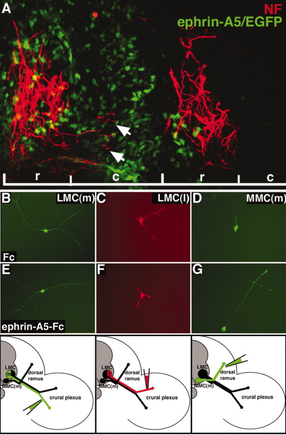
Neuron responses to ephrin-A5 are distinct. A, Sagittal section showing that NF+ MMC(m) axons (red) grow aberrantly (arrows) into the caudal half-sclerotome when the domain of ephrin-A5 expression (green) in the sclerotome is expanded. r, Rostral half-sclerotome; c, caudal half-sclerotome. B, E, LMC(m) neurons that are EphA4-demonstrate robust growth on ephrin-A5 or in controls (Fc). C, F, The growth of LMC(l) neurons that express EphA4 is reduced on ephrin-A5 substrates, compared with controls (C). D, G, MMC(m) neurons display poor growth in control cultures but exuberant neurite growth on ephrin-A5 substrates. Bottom, Schematic diagrams showing that retrograde labeling of the ventral nerve trunk to label LMC(m) neurons, of the dorsal nerve trunk to mark LMC(l) neurons, and of the dorsal ramus to label MMC(m) neurons is precise.