
Figure 7.
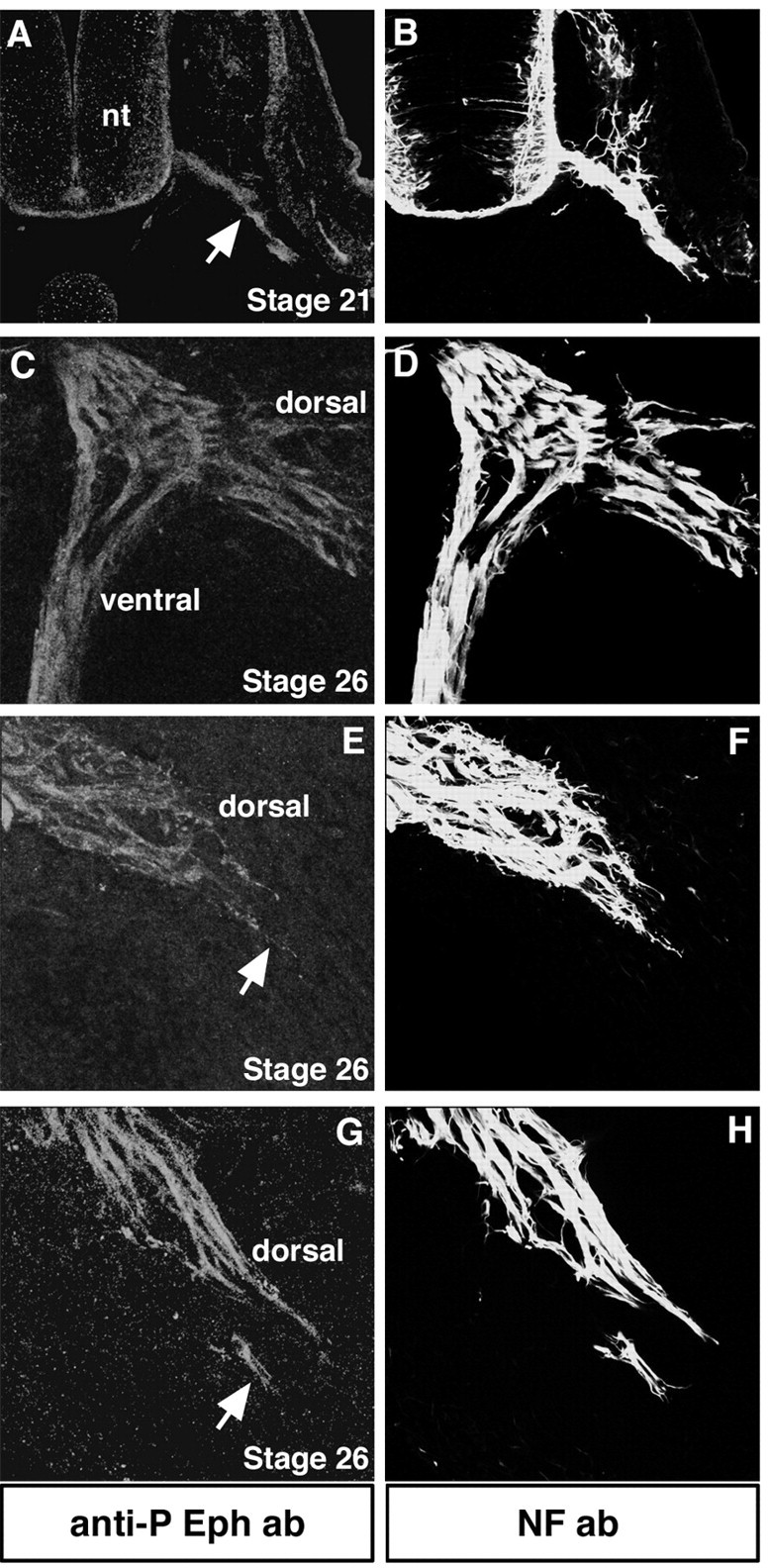
Eph phosphorylation localizes to the axon shafts and distal tips of LMC neurons in vivo. All transverse sections were labeled with anti-phosphorylated Eph antibody to detect Eph activation (red) and NF antibody to label all axons (green). A, B, At stage 21, before motor axons arrive to the base of the limb, they exhibit Eph phosphorylation (arrow). nt, Neural tube. C, D, At the level of the crural plexus at stage 26, dorsal and ventral projecting axons display Eph phosphorylation. E–H, Distal tips of LMC(l) axons (arrows) show Eph phosphorylation.