
Figure 6.
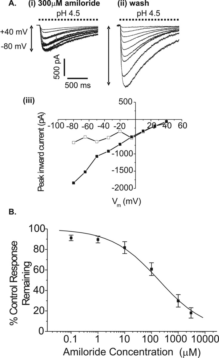
Effect of amiloride on the proton-gated current of rat retinal ganglion cells. A, pH 4.5-evoked currents recorded from a single voltage-clamped cell between –80 and +40 mV in 15 mV increments, in the presence (i) and absence (ii) of 300 μm amiloride. Aiii shows the current–voltage relationships from Ai (squares) and Aii (circles). B, Cells were voltage clamped at –80 mV, bathed in amiloride for 60 sec (0.1, 1, 10, 100, 1000, or 3000μm in random order), and multiple pH 4.5-evoked currents were recorded at 60 sec intervals until steady-state block was observed. Mean peak inward currents shown above (±SEM; n = 5–8 cells) have been normalized to amiloride-free control recorded just before every amiloride application. Sigmoidal curve parameters: IC 50, 188 ± 39 μm; slope, 0.49 ± 0.05.