
Figure 8.
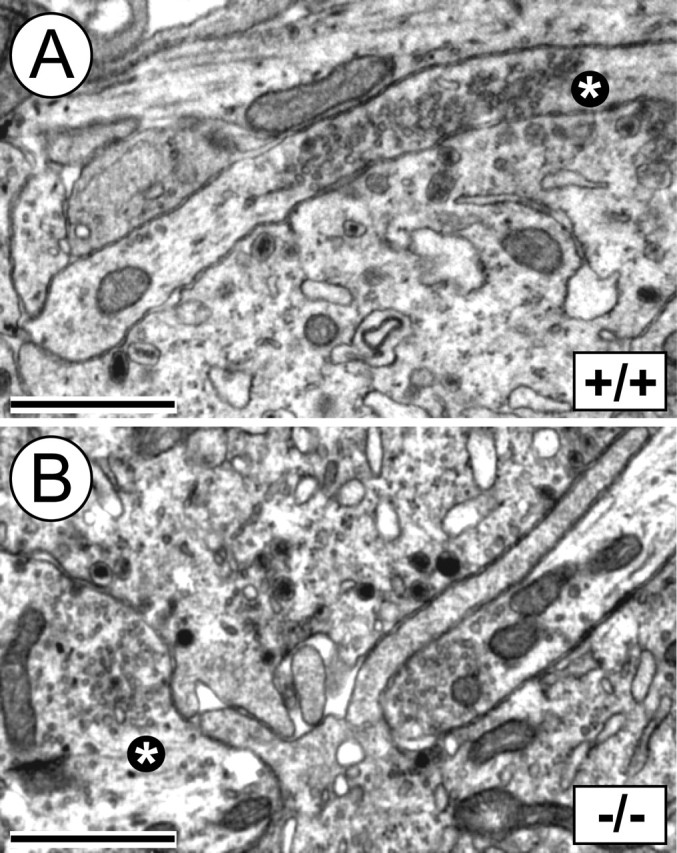
Electron microscopy of nerve terminals on adrenal chromaffin cells in E18.5 LIFRβ (+/+) and LIFRβ (−/−) mice. A, B, Axon terminals abutting chromaffin cells show clear synaptic vesicles and no overt morphological differences when comparing LIFRβ (+/+) (A) and LIFRβ (−/−) (B) adrenal medullae. The asterisks mark evaluated axonal terminals. Scale bars, 1 μm. Quantification of nerve terminals on chromaffin cells failed to reveal any significant difference between LIFRβ (−/−) mice and wild-type littermates. Six wild-type and eight LIFRβ knock-out mice were analyzed.