
Figure 5.
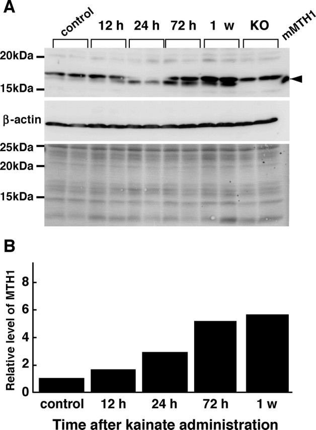
Increased expression of MTH1 protein in the hippocampus under the excitotoxicity. A, Western blotting analysis of MTH1 protein. Whole-cell extracts prepared from the mouse hippocampus prepared at 12, 24, 72 h, and 1 week after kainate administration were subjected to a Western blotting analysis with anti-MTH1 and anti-β-actin. The Western blotting results are shown in the top (MTH1) and middle (β-actin) panels. The bottom panel is a Ponceau S-stained filter before incubation with the antibodies to confirm sample loading. Extracts from MTH1-null mouse (KO) and recombinant mouse MTH1 (mMTH1) were also subjected to Western blotting. The arrowhead indicates mMTH1. B, The increased expression of MTH1 protein in the hippocampus after kainate administration. The amount of MTH1 protein determined by Western blotting was normalized by that of β-actin at each time point, and the mean value of the relative amount of MTH1 protein at each time point to that of the control is shown in the bar graph. n = 2 mice per group.