Figure 8.

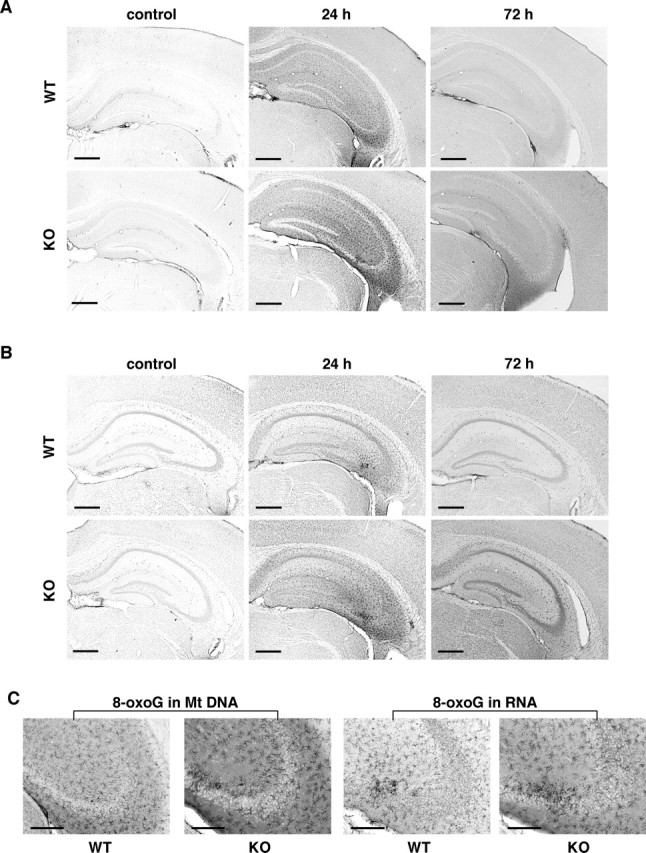
MTH1 deficiency augments the kainate-induced accumulation of 8-oxoG in the hippocampus. Wild-type (WT) and MTH1-null mice (KO) were injected with kainate (30 mg/kg, i.p.) or saline (control), and at 24 h and 72 h after kainate administration, brains were removed and coronal sections were subjected to IHC. A, The accumulation of 8-oxoG in mitochondrial DNA in the hippocampus. Free-floating sections treated with only RNase were subjected to IHC with N45.1 mAb. B, The accumulation of 8-oxoG in cellular RNA in the hippocampus. Free-floating sections without pretreatment were subjected to IHC with 15A3 mAb. C, The accumulation of 8-oxoG in microglia-like cells in CA3 under excitotoxicity. Magnified views (100×) of the CA3 subregions from A (24 h) [8-oxoG in mitochondrial (Mt) DNA] and B (24 h) (8-oxoG in RNA) are shown. Scale bars: A, B, 500 μm; C, 200 μm.
