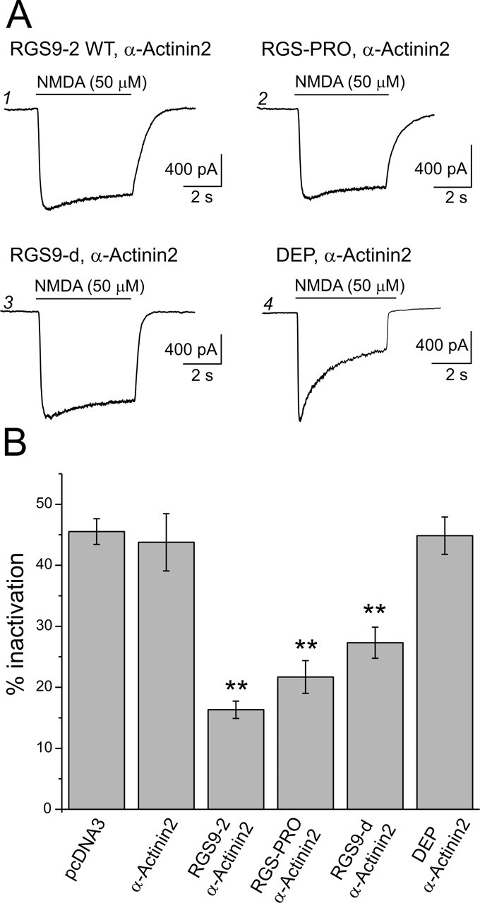
Figure 5.

The catalytic domain of RGS9-2 mediates the inhibition of CDI. A1, NMDAR current in a cell transfected with RGS9-2 and α-actinin-2 shows little inactivation, indicating a suppression of CDI. A2, A3, A similar suppression of CDI is seen in cells transfected with RGS-PRO and α-actinin-2 or RGS9-d (the catalytic domain of RGS9-2) and α-actinin-2. A4, In contrast, a cell transfected with the isolated DEP domain of RGS9-2 and α-actinin-2 exhibits CDI comparable with that seen in control cells. B, Summary plot comparing the magnitude of CDI under control conditions (pcDNA3 transfection, n = 10; α-actinin-2 and pcDNA3, n = 8) and that observed after transfection with RGS9-2 plus α-actinin-2 (n = 33), RGS-PRO plus α-actinin-2 (n = 12), RGS9-d plus α-actinin-2 (n = 21), and isolated DEP domain of RGS9-2 plus α-actinin-2 (n = 16). **p < 0.01 compared with α-actinin-2. Error bars indicate SE.
