
Fig. 1.
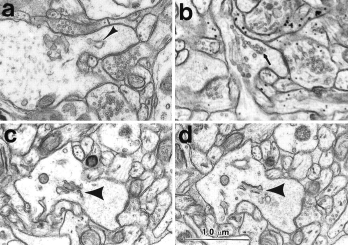
SER and spine apparatus in dendritic spines of hippocampal area CA1 at postnatal day 15. a, Cistern of SER (arrowhead) and b, vesicles of SER (arrow) in stubby dendritic spines. (This image is a bit soft, because it was enlarged from the edge of a negative that was photographed at 4K.) c, d, Adjacent serial sections through a spine apparatus (short thick arrow) in an emerging stubby-shaped dendritic spine with a perforated PSD on the spine head. It is likely that this spine represents the precursor of mushroom-shaped spines in the adults. Scale bar (shown in d): 1.0 μm.