
Fig. 3.
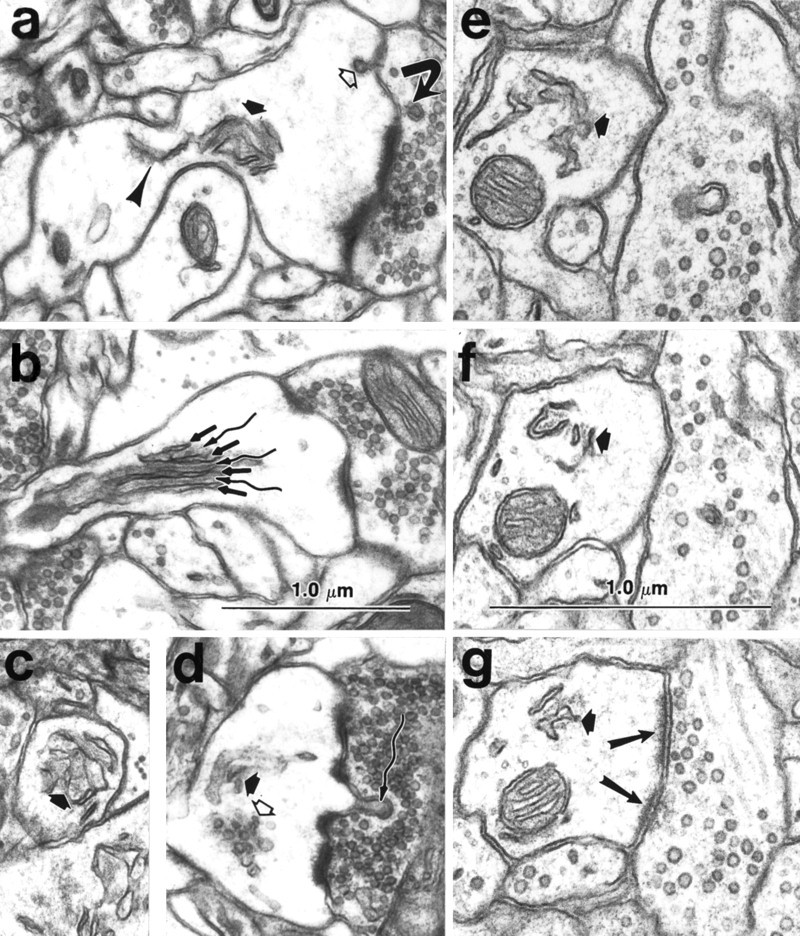
Mature dendritic spine apparatus.a, Spine apparatus (thick short arrow) elaborating from a single tubule of SER (arrowhead) in the dendritic shaft. A coated vesicle (open arrow) occurs at the membrane of the spine head, and a double-walled coated vesicle (curved arrow) occurs in the presynaptic bouton. See also Figure 5 for more discussion of this feature.b, Longitudinal section through a different spine apparatus clearly illustrating the lamination of SER (straight arrows) with dense staining plates (wavy arrows). c, Cross section of a spine apparatus (thick arrow) in a spine neck. d, Cluster of smooth vesicles (open arrow) in the head of a mushroom-shaped spine. This spine also contains a spine apparatus (thick filled arrow) and a spinule (wavy arrow) projecting toward the presynaptic bouton. A coated vesicle occurs in the presynaptic axon at the tip of the spinule (wavy arrow). e–g, Alternate serial sections through a spine apparatus (thick arrow) at the base of an emergent stubby dendritic spine that has a perforated PSD (two long arrows in g illustrate two portions of the PSD). In the dendritic shaft, microtubules surround the cross-sectioned mitochondrion clearly delimiting the cytoplasm of the shaft from the emerging spine, which has no microtubules, but instead contains only a web of filamentous material. Scale bars:a–g (shown in band f), 1.0 μm.