
Fig. 1.
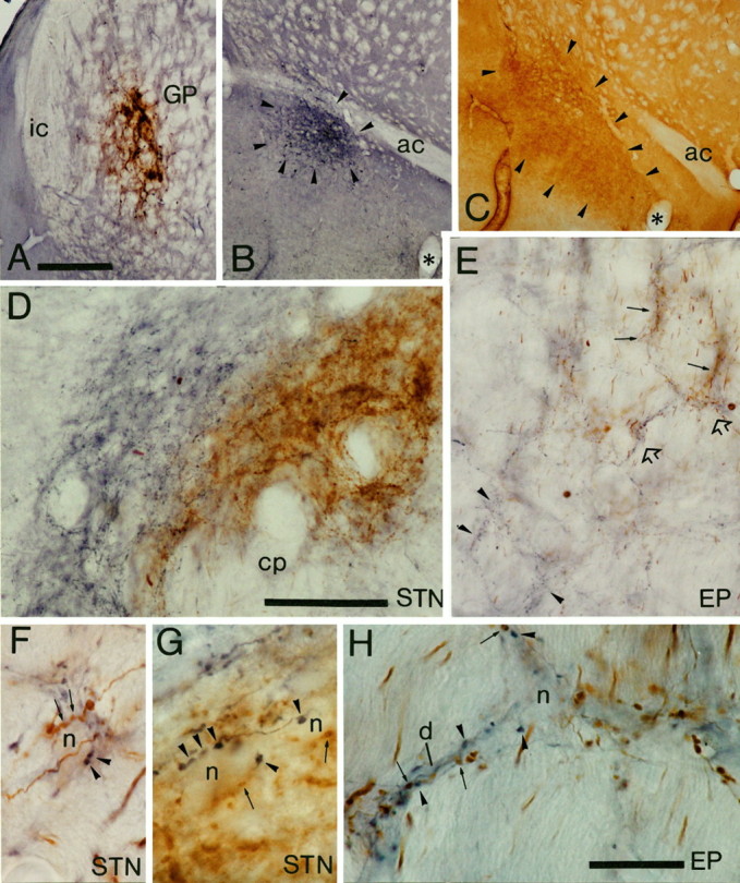
Light micrographs of anterograde labeling from the ventral pallidum and globus pallidus (GP) in the entopeduncular nucleus (EP) and subthalamic nucleus (STN). A–C, Sites of deposit of BDA in the globus pallidus (A) were revealed by using DAB as the chromogen, and PHA-L in the ventral pallidum (B) were revealed by using nickel-DAB as the chromogen for the peroxidase reaction. The extent of the tracer deposit in the ventral pallidum (arrowheads) was assessed by staining of adjacent sections to reveal substance P immunoreactivity (arrowheads) (C). D, Medium-power micrograph of anterograde labeling in the subthalamic nucleus. The fibers anterogradely labeled from the ventral pallidum (blue) occupy the medial and dorsal aspects of the STN, whereas those from the globus pallidus (brown) occupy the more lateral parts; however, the two sets of anterogradely labeled fibers are mixed at the interface between the projections. Note that the width of the fields of anterograde labeling is well within the dendritic diameter of subthalamic neurons. E, Medium-power micrograph of anterograde labeling in the entopeduncular nucleus. The fibers derived from the globus pallidus (brown; some indicated by arrows) and those derived from the ventral pallidum (blue; some indicated byarrowheads) are largely separate at this level, although there are areas of overlap of the two sets of fibers (some indicated byopen arrows and shown at higher magnification inH). F, G, High-power micrographs of unstained neuronal perikarya in the STN that are closely apposed by axonal swellings derived from both the ventral pallidum (blue; some indicated by arrowheads) and the globus pallidus (brown; some indicated byarrows). Individual perikarya are apposed by several boutons from each site. H, High-power micrograph of the region of the entopeduncular nucleus indicated by open arrows in E. An unstained neuronal perikaryon (n) and dendrite (d) are closely apposed by axonal swellings derived from both the ventral pallidum (blue; some indicated by arrowheads) and the globus pallidus (brown; some indicated byarrows). ac, Anterior commissure;cp, cerebral peduncle; ic, internal capsule. Scale bars: A–C (shown in A), 500 μm; D, E, (shown in D), 100 μm;F–H (shown in H), 20 μm.