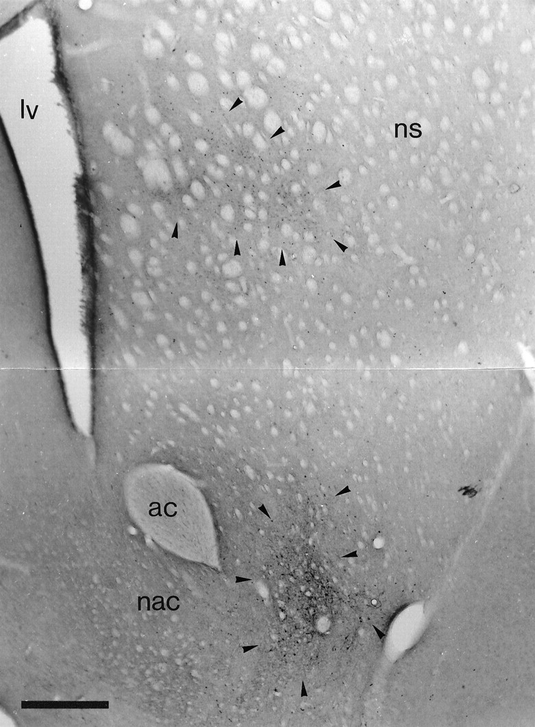
Fig. 3.

Retrograde labeling in the striatal complex after tracer deposits in the ventral pallidum and the globus pallidus. Low-power micrograph of the neostriatum and nucleus accumbens of the same animal as illustrated in Figure 2C. The section was incubated to reveal retrogradely transported PHA-L that was injected in the ventral pallidum and BDA that was injected in the globus pallidus. Although it is difficult to distinguish the labeling in this black and white micrograph, neurons retrogradely labeled from the globus pallidus (labeled with DAB; area indicated by arrowheads) are present only in the dorsal part of the neostriatum, whereas neurons retrogradely labeled from the ventral pallidum (labeled with Ni-DAB; area indicated by arrowheads) are present only in the most ventral aspects of the neostriatum and the nucleus accumbens. ac, Anterior commissure; lv, lateral ventricle; nac, nucleus accumbens;ns, neostriatum. Scale bar, 0.5 mm .
