Fig. 4.

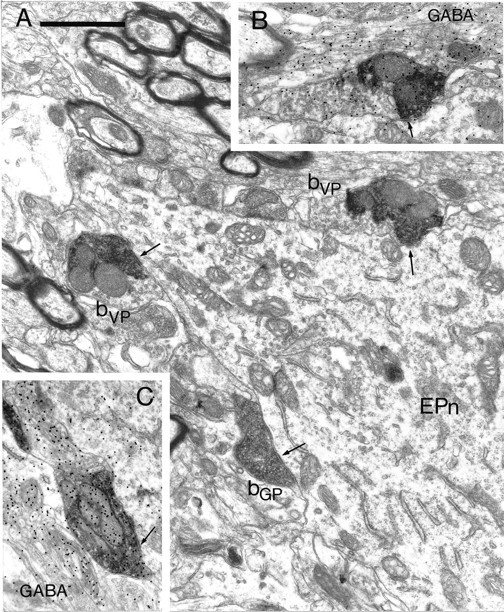
Synaptic convergence of terminals derived from different functional domains of the pallidal complex in the entopeduncular nucleus. A, Electron micrograph of part of a proximal dendrite of a neuron in the entopeduncular nucleus (EPn). The neuron is apposed by three anterogradely labeled boutons, each of which forms symmetrical synaptic contact with the neuron (arrows). Two of the boutons contain the BDHC peroxidase reaction product that was used to localize the PHA-L anterogradely transported from the ventral pallidum (bVP). The third bouton (bGP) contains the DAB reaction product that was used to localize the BDA anterogradely transported from the globus pallidus. Note that the BDHC reaction product that labels the terminals from the ventral pallidum has an irregular appearance and occupies only part of the labeled bouton, leaving many vesicles visible that do not have reaction product associated with them. In contrast, the DAB reaction product that labels the boutons from the globus pallidus is amorphous and occupies the whole of the labeled structure.B, A serial section of the upper of the two boutons derived from the ventral pallidum. This section was processed by the post-embedding immunogold method to reveal GABA immunoreactivity. The bouton has a high density of immunogold particles associated with it (index of GABA immunoreactivity = 3.79). C, Serial section of the bouton derived from the globus pallidus labeled by the post-embedding immunogold method to reveal GABA immunoreactivity. The bouton has a high density of immunogold particles associated with it (index of GABA immunoreactivity = 7.51). Scale bar (shown in A):A–C, 1 μm.
