Fig. 7.

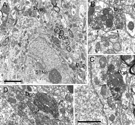
Synaptic convergence of terminals derived from different functional domains of the pallidal complex in the subthalamic nucleus. A, Part of the cell body of a neuron in the subthalamic nucleus (STNn) that is apposed by three anterogradely labeled terminals (bVP, bGP) shown at higher magnification inB–D. In this animal the injections were reversed, i.e., the PHA-L was injected in and anterogradely transported from the globus pallidus, and the BDA was injected in and anterogradely transported from the ventral pallidum. One of the boutons is lightly labeled with the DAB reaction product that adheres to vesicle and mitochondrial membranes, identifying it as arising in the ventral pallidum (bVP). It is shown at higher magnification inB. The bouton forms symmetrical synaptic contacts (arrows) with the subthalamic neuron. The other two boutons, shown at high magnification in C andD, are strongly labeled with the crystalline BDHC reaction product, which has an irregular appearance. These boutons are thus derived from the globus pallidus (bGP) and form symmetrical synaptic contacts with the neuron (arrows). Note that micrograph D is a different serial section of that shown in A. Scale bars:A, 2 μm; B–D (shown inC), 1 μm.
