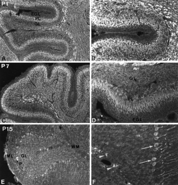
Fig. 3.

Evolution of PDGF-αR immunoreactivity in the developing cerebellum. Immunodetection of PDGF-αR in the cerebellum at P1 (A, B), P7 (C, D), and P15 (E, F). PDGF-αR immunoreactivity is detected in the Purkinje cell layer, the granule cell layer, and the presumptive white matter (A). High-magnification view showing PDGF-αR immunoreactivity on Purkinje and granule cells. Note the lack of expression in the external germinal layer (B). The expression of PDGF-αR is also widely distributed in the P7 cerebellum (C). High-magnification view of immunostained Purkinje and granule cells (D). At this period of development, the protein is well evidenced in the Purkinje cell soma and dendrites. At P15, PDGF-αR immunoreactivity is considerably decreased in the granule cell layer and is not detected on white matter fibers (E, F). The expression is always observed in the soma and dendritic tree of Purkinje cells (arrows inF) and in oligodendrocyte progenitors (arrowheads in F). Magnification:A, C, E, 100×; B, D, F, 200×.EGL, External germinal layer; ML, molecular layer; PL, Purkinje cell layer;GL, granule cell layer; WM, white matter.
