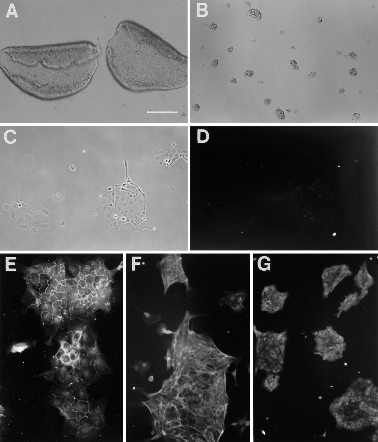
Fig. 1.
Utricular epithelial cell cultures and immunostainings. A, Two intact utricular epithelial sheets separated from P4–P5 rats. B, Partially dissociated epithelial sheets at the time of plating. C, D, Phase and fluorescence pictures of a 2 d epithelial cell culture labeled with an antibody against vimentin.E–G, Immunostaining of the 2 d cultures with an antibody against ZO1, a phalloidin-FITC conjugate, and an antibody against pan-cytokeratin, respectively. Scale bars: A, B, 200 μm; C–G, 100 μm.

