
Fig. 1.
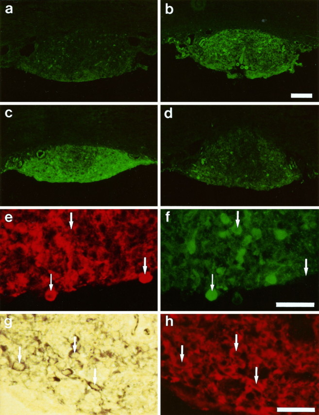
Immunocytochemical and NADPH–diaphorase staining of 10-μm-thick sections of the rat subfornical organ (SFO).a–d, Comparison of cGMP immunoreactivity after normal treatment (control, a) and after 10 min incubation with sodium nitroprusside (SNP; b),S-nitroso-N-acetyl-dl-penicillamine (SNAP; c), and 3-morpholinosydnonimine (SIN-1;d). Markedly increased levels of cGMP are visible after treatment with NO donors. Scale bar (shown in b): 200 μm. e, f, Double labeling of an identical SFO section for neuronal NO synthase (nNOS; e) and cGMP (f) immunoreactivity. One cell labeled by both antibodies is marked with a small arrow, whereas the large arrows mark cells exclusively stained by the cGMP antibody or nNOS antibody, indicating a codistribution rather than a colocalization. Scale bar (shown inf): 40 μm. g, h, Colocalization of NADPH–diaphorase staining (g) with nNOS immunoreactivity (h) in an identical section of the SFO. Examples of cells labeled by both techniques are marked with arrows. Scale bar (shown in h): 40 μm.