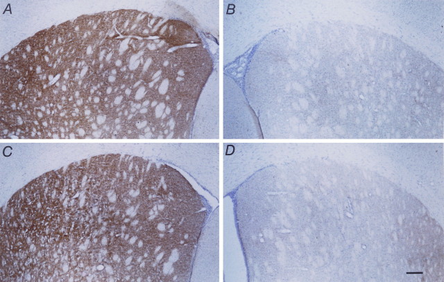
Fig. 4.
Micrographs showing TH-IR staining in the striatum 1 week after MFB axotomy in animals that received capsules containing either BHK-GDNF cells (A, B) or BHK control cells (C, D). A and C show the control (nonlesioned) side, whereas B andD show the lesioned side. Note the lack of TH-IR staining on the lesioned side in both cases, illustrating the absence of regrowth or sprouting of remaining fibers into the deinnervated striatum in either case. Scale bar, 200 μm.

