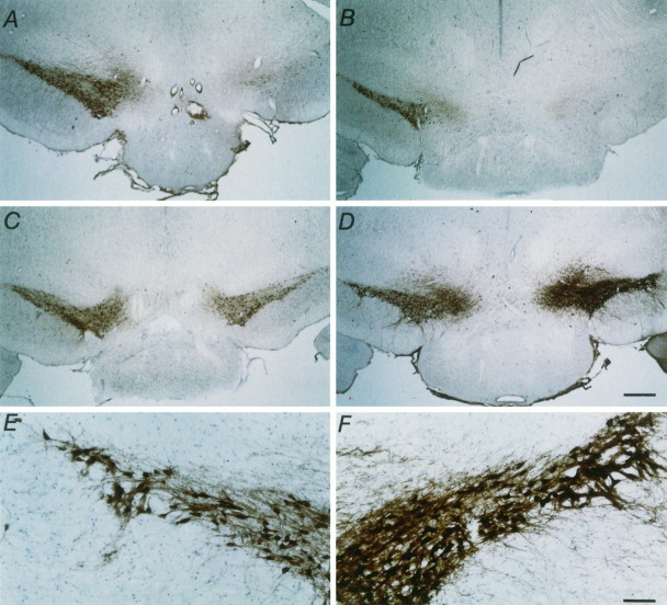
Fig. 6.
Micrographs showing TH-IR staining in the SN pars compacta 1 week after MFB axotomy. Low power pictures of coronal sections show the SN pars compacta. A, A typical control animal with no implant. B, A typical control animal that had received a capsule containing control BHK cells. C, D, Two typical animals that had received a capsule containing GDNF-secreting BHK cells. Note the almost total loss of TH-positive staining in the two control groups (A, B), whereas the group that received BHK-GDNF cells showed substantial rescue (C, D). Scale bar, 500 μm. High power photographs show TH-IR staining in the nonlesioned (E) and lesioned (F) SN pars compacta of the animal shown inD that had received a BHK-GDNF implant. Note the intense TH-IR staining and extensive dendritic sprouting in the lesioned SN pars compacta (F), as compared with the nonlesioned SN pars compacta (E). Scale bar, 100 μm.

