
Fig. 2.
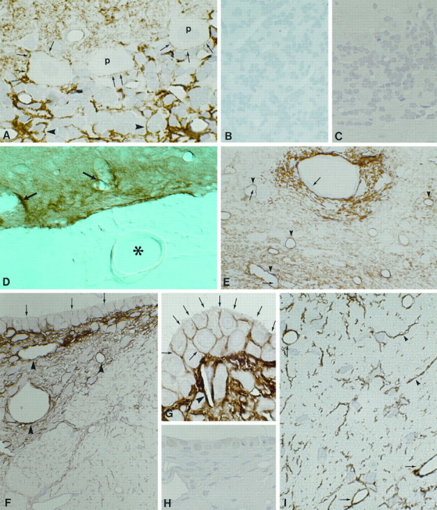
Immunocytochemical localization of AQP4 in rat brain. A, Cryosection of cerebellar cortex.Arrowheads and arrows indicate labeled glial processes in contact with granule cells and Purkinje cells. Magnification, 1000×. B, Control using affinity-purified antibody preabsorbed with excess immunizing peptide reveals no labeling. Magnification, 480×. C, Cryosection of cerebellum incubated with anti-AQP3 (Ecelbarger et al., 1995) demonstrates no labeling. Magnification, 480×. D, Vibratome section of the ventral brain surface at the level of the mesencephalon. Labeling is concentrated close to intracerebral vessels (arrows) and pia but is not associated with vessels in the subarachnoidal space (asterisk) or with arachnoid trabeculae. Magnification, 270×. E, Cryosection of thalamus demonstrates the predominant labeling of glial processes in the vicinity of vessels (arrowheads). Endothelial cells (arrows) and neurons are unlabeled. F, Cryosection of subfornical organ. Perivascular glial processes are heavily labeled (arrowheads). Distinct labeling of basolateral plasma membranes of ependymal cells is also present (arrows). Magnification, 480×. G, Higher magnification of basolateral labeling of ependymal cells (arrows) covering the subfornical organ and heavy labeling of perivascular glial cells (arrowhead). Magnification, 1000×. H, Immunolabeling control. Magnification, 480×. I, Cryosection from spinal cord reveals strong labeling of glial cells (arrowheads) and perivascular glial processes (arrows). Magnification, 480×.