
Fig. 7.
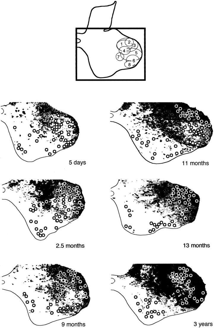
Corticospinal anterograde labeling in the gray matter at the C8–Th1 junction at different ages: 5 d, 2.5, 9, 11, and 13 months, and 3 years (case 3). The black frame at the top (from Fig. 9 in Jenny and Inukai, 1983) indicates the region of gray matter represented belowand also shows the distribution of selected hand muscle motor nuclei (for numbering, see Fig. 3). The corticospinal terminal labeling (inblack) and the location of motoneurons (circles) have been obtained from digitized paratungstate/TMB-reacted sections.