
Fig. 4.
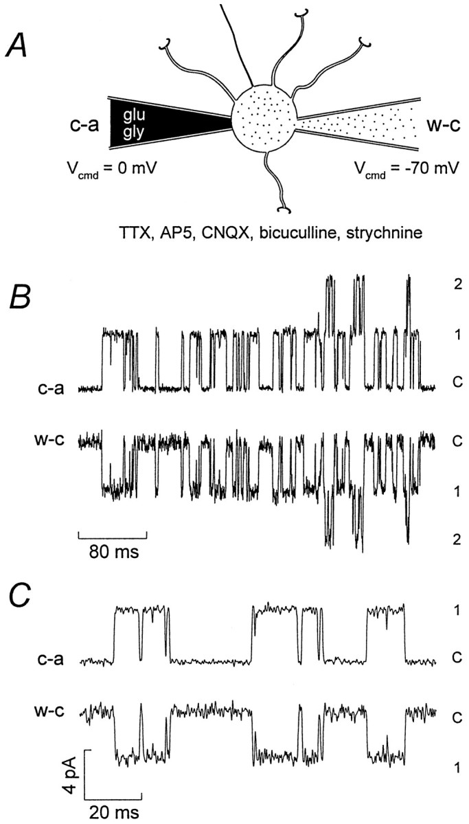
Isolation of extrasynaptic NMDA channel openings.A, Diagram showing the approach used to record selectively somatic NMDA channel openings. A granule cell with four dendrites and an axon is shown with cell-attached (c-a) and whole-cell (w-c) electrodes positioned on the soma. The diagram is not to scale; the mean soma diameter of granule cells (P9–P14) is ∼7 μm, and the dendrite length is ∼13 μm (M. Farrant, unpublished data; see also Silver et al., 1992).B, Paired current records from a single granule cell (P12). The top trace is from the cell-attached (c-a) electrode (Vcmd = 0 mV) with outward currents indicating the opening of 1 or2 channels from the closed level (C). The patch electrode contained 1 μm glutamate, 3 μm glycine, 10 μm bicuculline methobromide, 5 μm CNQX, and 200 nm strychnine. Thebottom trace is from the whole-cell (w-c) electrode (Vcmd = −70 mV), with inward currents mirroring those recorded from the cell-attached electrode. The bath solution contained 10 μm bicuculline methobromide, 5 μm CNQX, 10 μm AP5, 200 nmstrychnine, and 300 nm TTX. C, Currents from the same cell as B, shown on a faster time course. The current scale bar applies to both B andC. For display, the currents were digitized at 10 kHz after filtering at 1 kHz (8-pole Bessel, −3 dB).