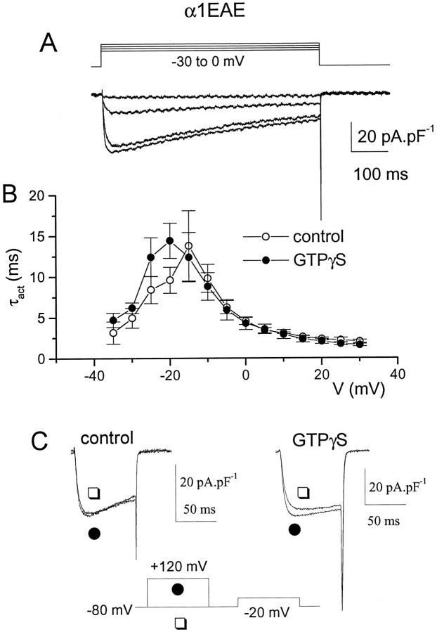
Fig. 6.
Cells were transfected with the α1EAE chimera, together with α2-δ and β1b, and IBawas recorded after 3 d in culture. A,IBa was activated by 600 msec steps to examine the rate of inactivation of α1EAE. B,IBa was activated by 100 msec steps, and τact was measured as described in the legend to Figure 3for cells recorded in the presence of 100 μm GTPγS in the patch pipette (•, n = 5), or in its absence (○, n = 7). C,IBa was recorded in the presence (•) or absence (□) of a +120 mV depolarizing prepulse applied 30 msec before the test pulse to −20 mV for a control cell (left) and a cell containing GTPγS (right). Prepulse facilitation was observed only in the GTPγS-containing cell.

