
Fig. 5.
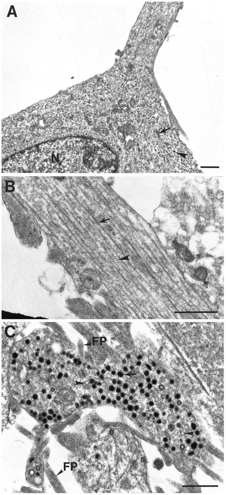
Electron micrographs of differentiated CAD cells.A, Cell body and initial segment of a process in differentiated CAD cells. The cytoplasm contains ribosomes (arrowhead), mitochondria (asterisk), rough endoplasmic reticulum (arrow), and microtubules.B, A typical process is filled with parallel microtubules (arrow) and intermediate filaments (arrowhead). C, Dense-core vesicles (arrow) and clear vesicles (arrowhead) in terminal. FP, Filopodia; N, nucleus. Scale bars, 1 μm.