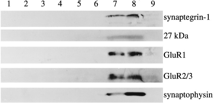
Fig. 9.
Comigration of RGDS-binding proteins and synaptic markers across density gradients. Fresh forebrain P2 suspensions were applied to 3–25% Percoll gradients to isolate synaptosomes, as described in Materials and Methods. The interfacial zones of the gradients were separated, washed by centrifugation, and hyposmotically treated. Aliquots of the soluble fraction from interfacial zones 2–5 were concentrated and prepared for immunoblotting (lanes 1–4, respectively); lysed membranes from zones 1–5 (lanes 5–9; 70 μg protein each) were also immunoblotted. The following antibodies were used: anti-β1 (labeled 55 kDa synaptegrin-1), anti-αvβ3 (labeled a 27 kDa antigen), anti-GluR1, anti-GluR2/3, and anti-synaptophysin.

