
Fig. 4.
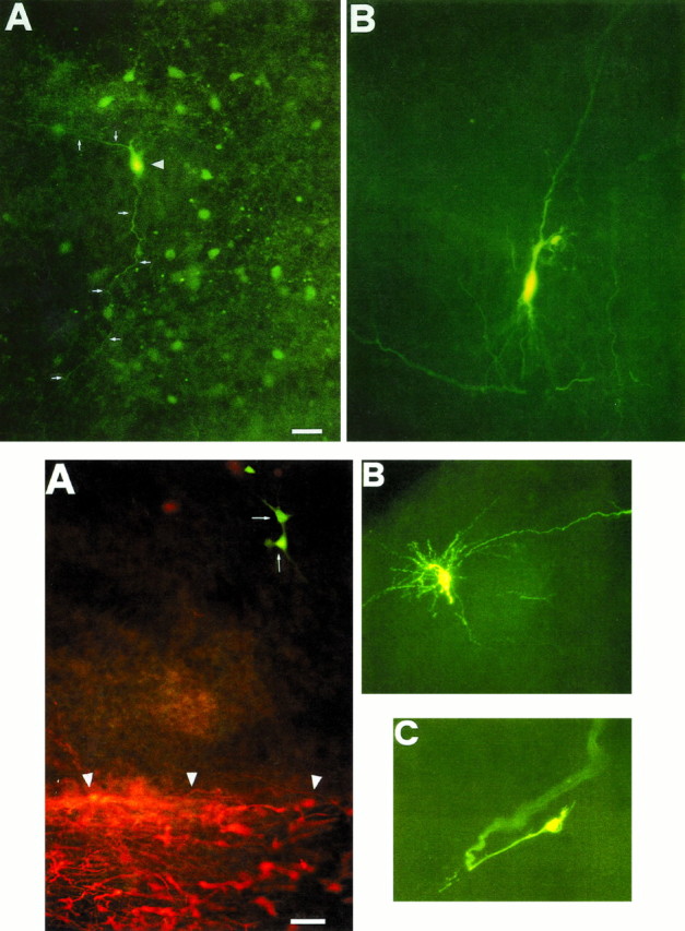
Top. Biocytin-injected hilar interneuron, but not pyramidal cell, reveals surrounding cells. A, P3 hilar cells in a 50 μm hippocampal slice. The injected interneuron (arrowhead) is coupled to a cluster of surrounding cells. Two neuronal processes (white arrows) connect neighboring cells. B, Staining of a single CA3 pyramidal neuron. Scale bar, 44 μm.