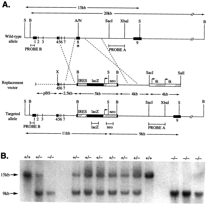
Fig. 1.
GABAA receptor α6subunit gene disruption by homologous recombination. A, Wild-type α6 gene, targeting (replacement) vector, and the disrupted α6 gene structures. Numbersindicate exons. On the Replacement vector, thebroken line indicates pBluescript (pBS) sequences. On the wild-type allele, theasterisk marks exon 8 where the IRES lacZ/neo cassette was inserted. Only relevant restriction sites are shown. A, AflII;B, BamHI; N,NcoI; S, SphI;X, XhoI. Arrows mark theneo and tk gene promoter sites and direction of transcription. The lacZ coding sequence orientation is the same as the α6 gene, thus permitting its translation from the IRES sequence (striped box) to be initiated on the mRNA derived from the α6 promoter. Expected restriction fragment lengths diagnostic for homologous recombination and the probes used to detect these are marked by double-headed arrows andhorizontal bars, respectively. B, Confirmation of α6 mutant allele germline transmission. Biopsy tail DNA samples were digested with SphI, electrophoresed, and Southern-blotted. The membrane was probed withPROBE A (3′ flanking). Wild-type (+/+) individuals give a 15 kb band, the homozygous null (−/−) animals give a 9 kb band, and heterozygotes (+/−) give both bands.

