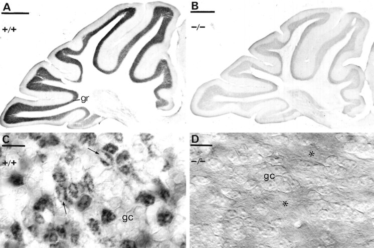
Fig. 7.
Immunodetection of the δ subunit of the GABAA receptor in α6 +/+ (A,C) or α6 −/− (B, D) cerebella, using a polyclonal antibody δ R7 and immunoperoxidase reaction. The granule cell layer showed intense immunoreactivity in α6 +/+ animals but almost no staining was observed in the α6 −/− mouse. C, At higher magnification, it is evident that the δ subunit is localized mainly in the glomeruli, granule cell bodies (gc) being only weakly outlined. The glomeruli appear as dark rings of granule cell dendrites surrounding a pale center (arrows) representing the unstained mossy fiber terminal. D, In the α6 −/− mice, both the granule cell bodies (gc) and the glomeruli (asterisks) are immunonegative for the δ subunit. C andD were photographed using DIC optics. Scale bars:A, B, 500 μm; C, D, 10 μm.

