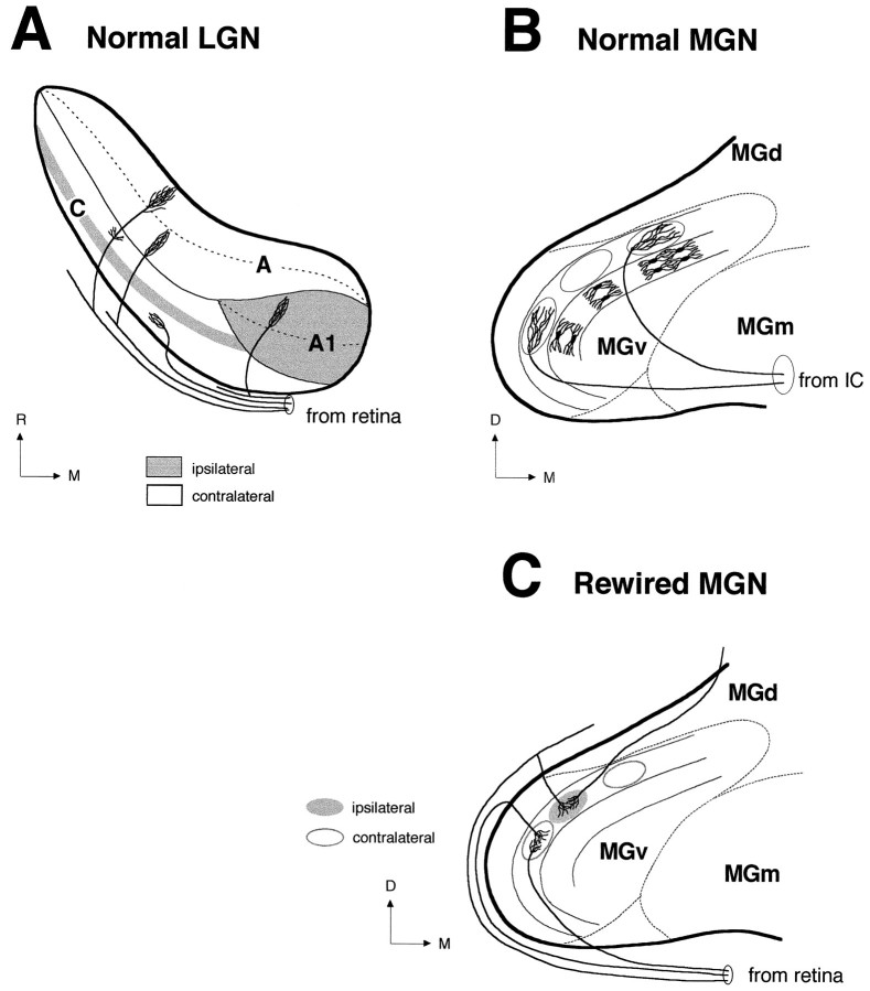
Fig. 12.
Terminal patterns of afferent projections to the normal LGN, normal MGv, and rewired MGv. A, Schematic representation of retinal projections to the normal ferret LGN in the horizontal plane. Projections from the ipsilateral (gray areas) and contralateral (empty areas) retinae segregate into eye-specific layers in the LGN (A, A1, C) (Linden et al., 1981). Within layers A and A1, afferent projections from the contralateral and ipsilateral eyes, respectively, further segregate into on-center and off-center sublayers (dashed lines) (Hahm et al., 1991). Three main morphological types of retinogeniculate axons have been described in the ferret LGN (Roe et al., 1989; Pallas et al., 1994): X axons project to the A layers, Y axons to the A and C layers, and W axons only to the C layers. B, Schematic representation of afferent projections from the IC to the normal MGv in the coronal plane. In MGv, projections from the IC form terminal clusters (ovals) aligned within dorsoventrally oriented fibrodendritic lamellae (Kudo and Niimi, 1980). The laminar pattern in MGv results from the ordered alignment of relay neurons (Morest, 1964, 1965; Winer, 1992). The position of the dendritic trees of these cells within a MGv lamina is illustrated (medial lamina). Axon arbors from the IC contribute to the laminar pattern of MGv by being elongated dorsoventrally (Morest, 1965) and anteroposteriorly (Pallas and Sur, 1994). C, Schematic representation of the novel retinal projection to MGv in the coronal plane. In rewired MGv, similar to the normal IC-to-MGv projection (B), retinal axons form terminal clusters aligned along lamellae. The lamellar organization of MGv is preserved in rewired ferrets (Pallas et al., 1990). However, similar to the normal retino-LGN projection (A), projections from the ipsilateral (gray oval) and contralateral (empty ovals) retinae are segregated in MGv. Thus, segregation of retinal afferents occurs in the form of adjacent but nonoverlapping eye-specific clusters. Clusters are formed by the convergence and overlap of several axon arbors. Retino-MGN arbors are more restricted than IC-to-MGN arbors (compare C andB) (Pallas and Sur, 1994) and resemble in size W-cell axon arbors in the LGN and SC (Pallas et al., 1994). A,A1, C, Layers A, A1, and C of the LGN;D, dorsal; M, medial; R, rostral.

