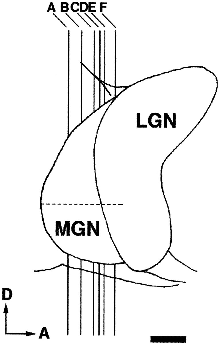
Fig. 2.

Lateral view of the dorsal thalamus. Thevertical lines (A–F) indicate the approximate anteroposterior level of each MGN section shown in Figure 1. The horizontal dashed line marks the rostrocaudal extent of the MGN. D, Dorsal; A, anterior. Scale bar, 1 mm.
