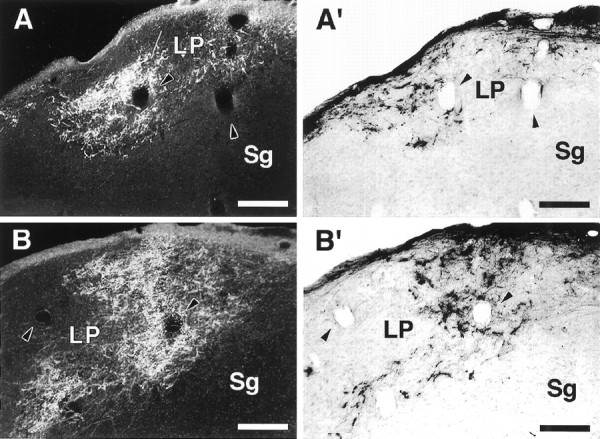
Fig. 5.

Retinal inputs to the LP in an adult rewired ferret. A, B, Dark-field micrographs of retino-LP projections labeled by an injection of WGA-HRP in the contralateral eye. A′, B′, Bright-field micrographs of ipsilateral retino-LPprojections labeled with CTB. Sections in A andB are immediately adjacent to sections inA′ and B′, respectively.Arrowheads point to corresponding blood vessels in adjacent sections. A composite drawing of A andA′ is shown in Figure 7. Note the terminal slab-like pattern of retinal projections to LP. Dorsal isup; medial is to the right. Scale bars, 200 μm.
