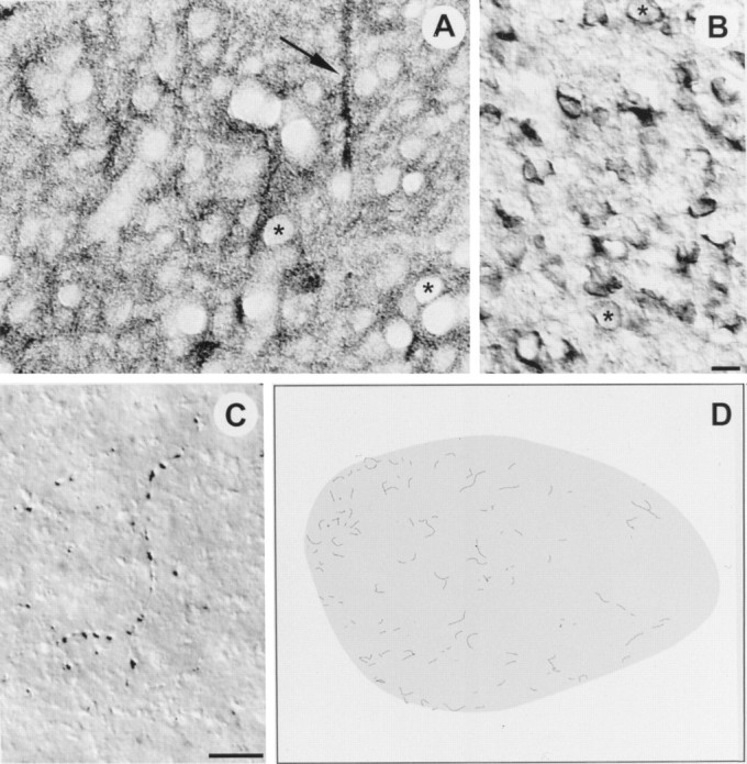
Fig. 5.

High-power digitized videomicrographs show immunoreactivity against the extracellular domains of TrkB (A), p75 (B), and TrkC (C) in RA of a juvenile male zebra finch; dorsal is up and medial is left in all images.A, TrkB immunoreactivity was found throughout RA. Fiber bundles (arrow), neuropil, and somata were all labeled by the antibody; only nuclei were left unstained, giving the tissue a punctuate appearance (asterisks show two examples of nuclei). The labeled fiber bundles (arrow) were oriented dorsoventrally, and they appeared to pass completely through RA, perhaps projecting to a target ventral to RA. These fiber bundles do not appear to be the axons of lMAN neurons, because lMAN axons enter RA dorsolaterally and laterally (Johnson et al., 1995). B, Immunoreactivity for p75 was found throughout RA and was localized primarily to neuronal somata, although fine labeled processes were sometimes observed. The asterisks show two examples of neuronal nuclei for comparison with nuclei in A.C, Antibody against TrkC only labeled fine processes in RA that seemed to be axons; these fibers often appeared to terminate by encircling somata within RA. D, The distribution of TrkC fiber labeling in RA based in a low-power camera lucida drawing. Note the TrkC labeling in RA was generally sparse but with a somewhat higher incidence of labeled fibers in medial RA. Scale bars (B,C), 10 μm.
