
Fig. 7.
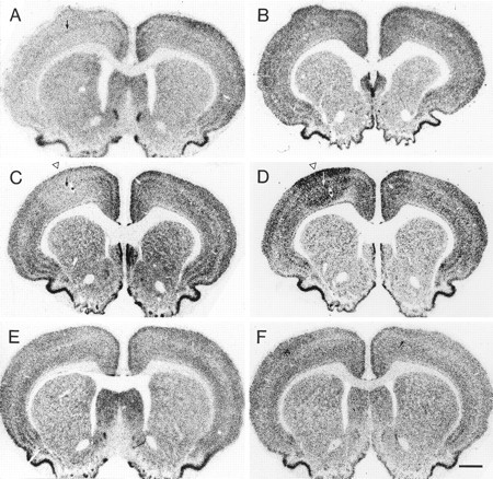
Photomicrographs showing in situhybridization histochemistry for GluR2 (left) and NR1 (right) mRNAs in frontal sections at the level of the motor cortex. A, In a rat that was injected with 35 ng TT and survived for 7 d, GluR2 mRNA levels were decreased throughout most neocortical areas on the injected side (left), whereas NR1 mRNA levels (B) were not clearly affected. C, In another rat that was injected with 35 ng TT and survived for 14 d, GluR2 mRNA levels were downregulated focally at the injection site in the motor cortex (open arrowhead), and NR1 mRNA levels (D) were focally upregulated at the injection site in the motor cortex (open arrowhead). Note the zone of NR1 mRNA downregulation around the focus of increased NR1 mRNA levels. E and F show that GluR2 and NR1 mRNA levels were not obviously changed in a saline-injected control rat. Arrows in A, C, andD indicate the injection site. Asterisksin C and D indicate the same blood vessel marked in Figure 1A. Scale bar, 1 mm.