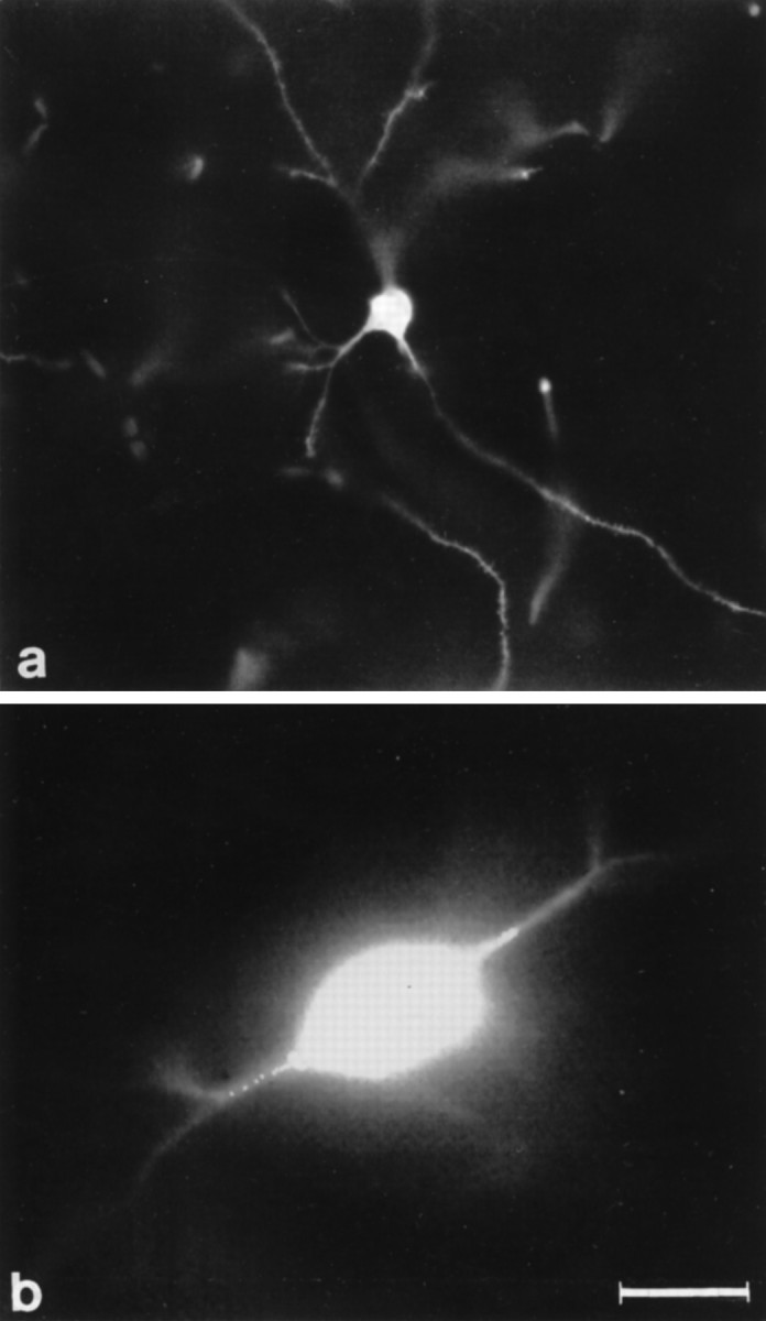
Fig. 1.

Morphological identification of striatal neurons. The figure represents two striatal neuronal subtypes: a biocytin-injected spiny striatal neuron (a) and a large LA interneuron filled with the dye fura-2 (b) (380 nm image, average of 256 frames) (for details, see Materials and Methods). Scale bar (shown in b): a, 70 μm;b, 30 μm.
