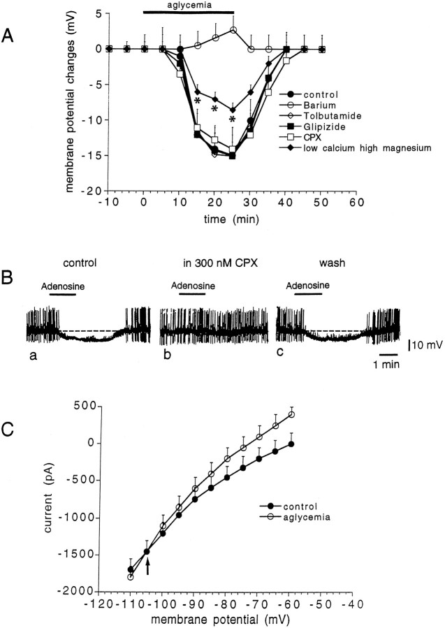
Fig. 8.
The aglycemia-induced hyperpolarization/outward current in LA interneurons is mediated by a K+ conductance.A, Time course of the membrane changes induced by 25 min of glucose deprivation in different experimental conditions: control (n = 9; filled circles), 300 μm barium (n = 4; open circles), 1 mm tolbutamide (n = 3; open rhombs), 100 nm glipizide (n = 3; filled squares), 300 nm CPX (n = 4; open squares), and low calcium (0.5 mm)/high magnesium (10 mm) solutions (n = 4; filled rhombs). Asterisks indicate significant difference from control values (p < 0.01).B, In an LA interneuron, bath application of 30 μm adenosine induced a membrane hyperpolarization (a); this membrane hyperpolarization was fully blocked by 300 nm CPX, an adenosine A1 receptor antagonist (b); after the washout of this antagonist, the hyperpolarizing action of adenosine was restored (c).C, The reversal potential of the aglycemia-induced outward current is indicated by the arrow (−105 mV;n = 4). This value was calculated by measuring the steady-state currents generated by long-lasting (1–3 sec) voltage steps of progressively increasing amplitude from the holding potential (−60 mV; n = 4) before (filled circles) and during glucose deprivation (25 min, open circles).

