
Figure 3.
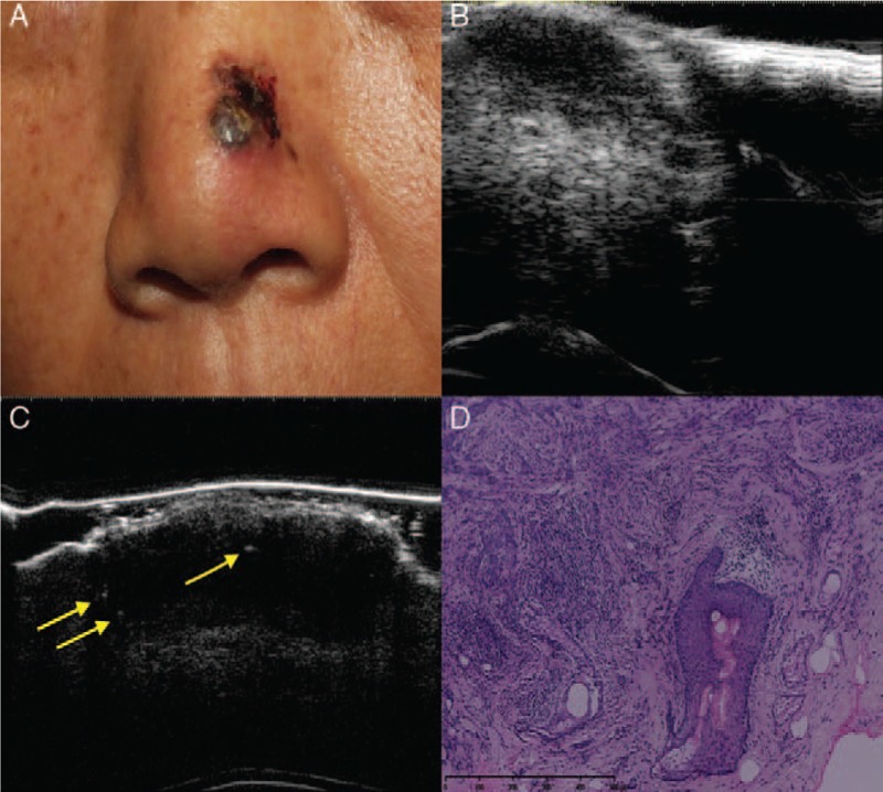
Basosquamous cell carcinoma. Clinical (A), ultrasonographic (B, C), and histologic (D) images of a basosquamous carcinoma lesion (high-risk sub-type). (A) The clinical image shows an ulcerated, pigmented, and nodular lesion on the left wing of the nose. (B) A 20-MHz ultrasound examination showed an ill-defined, irregular, hypo-echoic lesion. (C) A 50-MHz ultrasound examination showed epidermal and dermal thickening with epidermal irregularities and a hypo-echoic dermal lesion that presented three hyper-echoic spots (arrows). (D) The histology results indicated basosquamous cell carcinoma (hematoxylin-eosin staining, original magnification ×200).