
Fig. 5.
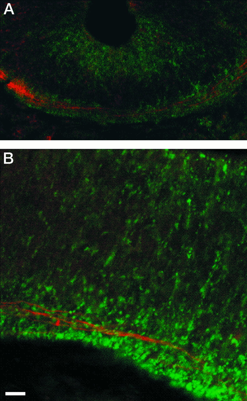
Double-labeling of netrin-1 and commissural fibers. Netrin-1 IR (green) is located around pioneering axons (red) that are either crossing the floor plate in the spinal cord (A) or in the mesencephalon (B, with midline slightly beyond the right edge of the photomicrograph). Little if any netrin-1 IR is present on the axons (areas with both green and redsignals are yellow). Transverse sections of stage 20 embryos were simultaneously processed with N1D and the β-tubulin antibody. Fluorescently tagged secondary antibodies were visualized by confocal microscopy. Scale bar: A, 7 μm;B, 5 μm.