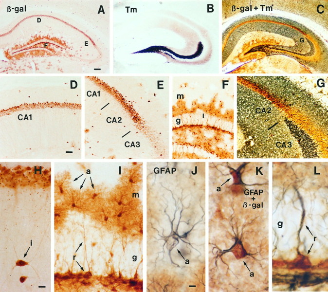
Fig. 5.

Immunohistochemical staining for β-galactosidase (β-gal) in transgenic mice expressing lacZ from the Lmo1-promoter 1.A, Survey of hippocampus stained for β-gal.B, Survey of hippocampus stained by the Timm method to show zinc-containing fibers, particularly in mossy fibers projecting from the dentate gyrus to CA3. C, Survey of hippocampus stained by both the Timm method and immunohistochemistry for β-gal illustrating the sharp border between Timm-stained fibers in CA3 and β-gal-positive neurons in CA2. D, E, Details ofA showing β-gal-positive pyramidal cells in CA1 and CA2 but not CA3, as well as scattered interneurons. F,Detail of A showing numerous β-gal-positive astrocytes in the dentate gyrus molecular layer (m) and hilus, and the absence of staining in granule layer (g) neurons. G, Detail ofC showing that β-gal-positive pyramidal neurons are present in CA2 but not in CA3 as delineated by the CA2–CA3 border demarcated by Timm-stained mossy fibers in CA3. H,Detail showing two β-gal-positive interneurons (i) in the stratum radiatum below CA1 pyramidal neurons. I,Detail of F showing the morphology of β-gal-positive astrocytes (a) in the molecular layer of the dentate gyrus. Note that in addition to the darkly stained cell bodies, the finely branched processes of the astrocytes are also stained and radiate out from the cell bodies to cover a large area and give the cells a tufted appearance (a). Not all astrocytes in the molecular layer are β-gal positive. Note also the β-gal-positive radial astrocytes (r) whose processes span the granule layer (g), and that the granule neurons are not stained. J, Detail showing an astrocyte (a) immunohistochemically stained only for GFAP using a blue chromogen. Note that blue staining for GFAP is present in the astrocyte processes and outlines but does not fill the cell body. This cell is located in a portion of the molecular layer similar to that shown in I, in between astrocytes stained positively for both β-gal and GFAP, as in K. K, Two astrocytes (a) located in the molecular layer that are immunohistochemically double-stained for both GFAP (blue) and β-gal (brown). Note that thebrown-stained cell bodies (β-gal) and dark blue-stained processes (GFAP) clearly belong to the same cells and compare in appearance with the single-stained astrocytes in I and J. L,Radial astrocyte (r) located in the granule layer that is immunohistochemically double-stained for both GFAP (blue) and β-gal (brown). Note that thebrown-stained cell body (β-gal) and dark blue-stained process (GFAP) clearly belong to the same cell, which compares in appearance with the single-stained radial astrocytes in I. Scale bars: A–C, 180 μm;D–G, 70 μm; H, I, 10 μm;J–L, 5 μm.
