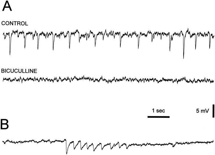
Fig. 12.
Spontaneous synaptic activity in kitten dLGN neurons. A, Voltage recordings show the presence of spontaneous hyperpolarizing potentials in a P3 neuron. These potentials were blocked by application of 50 μm bicuculline to the perfusion medium. B, Voltage recording from another P3 neuron shows the presence of a burst (4 Hz) of inhibitory postsynaptic potentials, similar in waveform but not in frequency to the sleep spindles (Steriade et al., 1993). Membrane potential was −55 mV inA and B.

