Fig. 2.

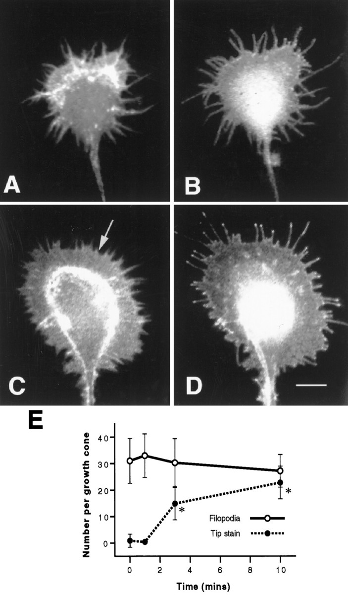
Accumulation of β1 integrin at filopodial tips is not dependent on the extension of filopodia. A, B, Representative growth cone that had been cultured in serum-free conditions, starved of NGF, and double-stained for F-actin (A) and β1 integrin (B). Filopodia are present, and β1 integrin is distributed evenly along their length. C, D, Representative growth cone cultured as in A and B, treated with 100 ng/ml NGF for 10 min, and double-stained for F-actin (C) and β1 integrin (D). Numerous veils have extended (C, arrow), and filopodia show accumulation of β1 integrin at their tips. Scale bar in D, 10 μm.E, Quantitation of the accumulation of β1 integrin at filopodial tips. Although the number of filopodia per growth cone remains constant, the number of tips with concentrations of β1 integrin increases after treatment with NGF. Results are representative of three separate experiments (n = 100). Error bars show SDs. Asterisks denote those values that are significantly different (p < 0.005) from control (0 min) values for tip staining.
