Fig. 4.
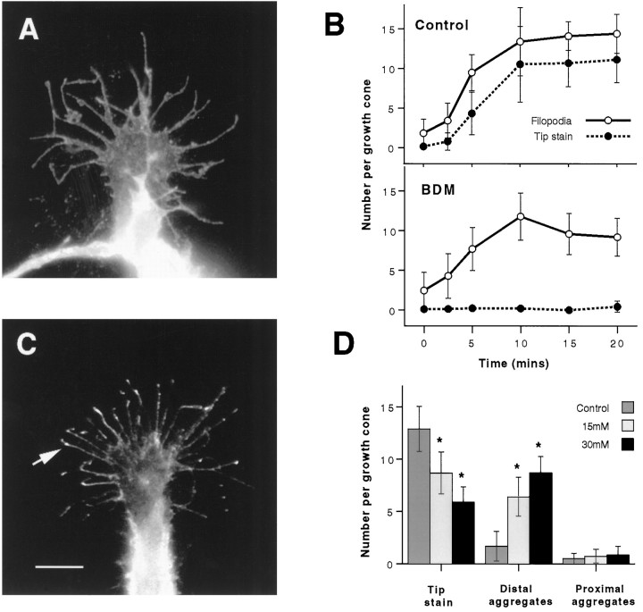
Treatment of cells with 15 mm BDM for 5 min before the addition of 100 ng/ml NGF (A,B) blocks the accumulation of β1 integrin at filopodial tips. A, A representative growth cone treated as above and stained for β1 integrin has numerous filopodia but no accumulation of β1 integrin at filopodial tips. B, Counts of filopodia and tip aggregates per growth cone demonstrate that, after treatment with BDM (as in A), the accumulation of β1 integrin at filopodial tips is blocked, but the formation of filopodia is only modestly reduced compared with parallel control cultures (as shown). Error bars show SDs. Results are representative of three separate experiments (n = 100). C, β1 Integrin aggregates remain either at the tips or in the distal halves of filopodia when 15 mm BDM is added to cells in which β1 integrin had previously accumulated at tips in response to 100 ng/ml NGF. A typical growth cone shows that the location of most β1 integrin aggregates is either at or just behind the tip (arrow) 5 min after the addition of BDM. Scale bar, 10 μm. D, Counts of β1 integrin aggregates at either the tip or in the distal (excluding the tip) and proximal halves of filopodia show that, after 5 min of treatment, although BDM causes the number of tip aggregates to decrease, most remain in the distal half of filopodia. Results are representative of three separate experiments (n = 25). Error bars show SDs.Asterisks denote those values that are significantly different (p < 0.005) from control (0 mm) values.

