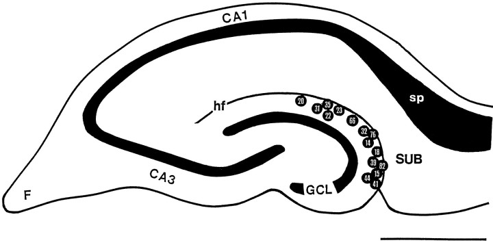
Fig. 1.
Location of somata of interneurons in the OML projecting to the subiculum. Schematic drawing of a transverse hippocampal slice shows the distribution of recorded and anatomically analyzed neurons. Internal reference numbers are shown within circles. Because of the overlap in the position of the somata, two neurons were omitted. Note that subiculum-projecting neurons could be found throughout the entire OML. CA1,CA3, Hippocampal regions CA1 and CA3; F, fimbria; GCL, granule cell layer; hf, hippocampal fissure; sp, stratum pyramidale of the subiculum; SUB, subiculum. Scale bar, 1 mm.

