Fig. 4.

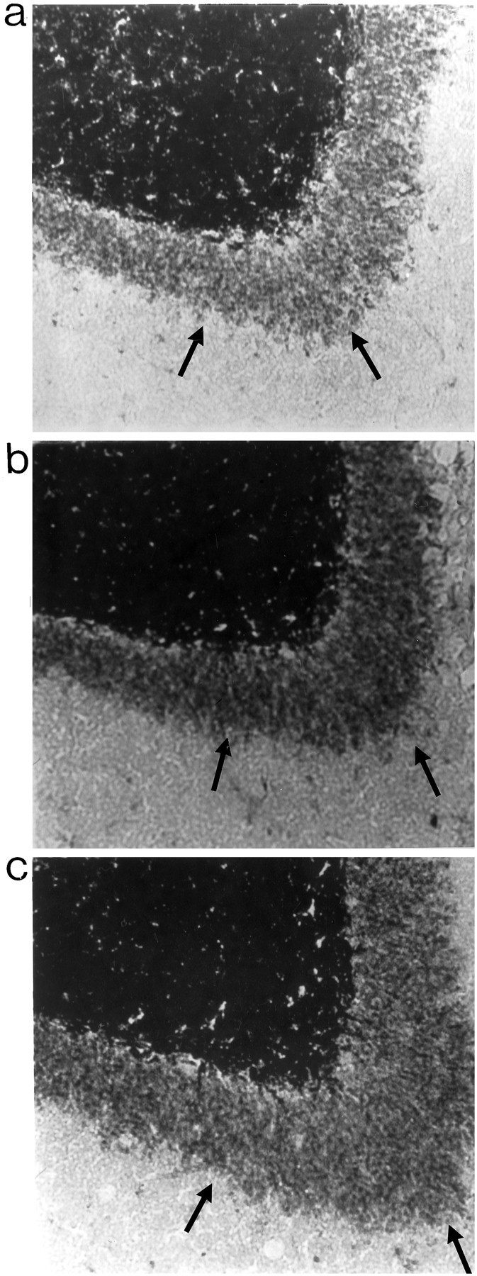
Timm staining in IML region. A,Representative examples of IML region of a nonkindled PBS-infused rat (a), a kindled PBS-infused rat (b), and a kindled NGF-infused rat (c). Arrows point to Timm granules in the IML region.

Timm staining in IML region. A,Representative examples of IML region of a nonkindled PBS-infused rat (a), a kindled PBS-infused rat (b), and a kindled NGF-infused rat (c). Arrows point to Timm granules in the IML region.