Fig. 1.

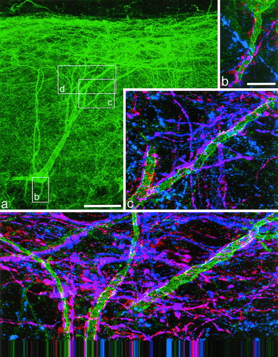
An NK1 receptor-immunoreactive neuron with the cell body 290 μm below the dorsal white matter, which received numerous contacts from substance P-immunoreactive varicosities. This cell is number 10 in Figure 3 and Table 1. NK1 receptor immunoreactivity is shown in green(a–d), substance P immunoreactivity inblue (b–d), and CGRP immunoreactivity inred (b–d). a, A low-magnification view of the cell body and dendritic tree revealed with antibody to the NK1 receptor. The boxes show the regions represented in b–d. b–d, Three regions of the dendritic tree shown at higher magnification. In each region axons with only substance P (blue) or CGRP (red) immunoreactivity are visible, but many show both types of peptide immunoreactivity and appear purple. Many varicosities with both substance P and CGRP immunoreactivity are in contact with the dendrites of the NK1 receptor-immunoreactive neuron; where these overlap with the NK1 receptor immunoreactivity, this appears white. a was obtained from 35 optical sections 1 μm apart, b and dfrom four and five sections, respectively, at 0.7 μm, andc from nine sections at 0.5 μm apart. Scale bars:a, 65 μm; b–d, 10 μm.
