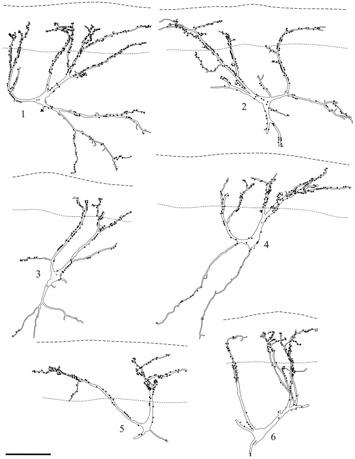
Fig. 2.
Drawings of 6 of the 12 NK1 receptor-immunoreactive neurons for which a detailed analysis of the contacts from substance P-immunoreactive varicosities was performed. The cells are all from parasagittal sections. In each case theupper dashed line represents the border between lamina I and the dorsal white matter, whereas the lower lineindicates the ventral limit of the dense plexus of peptide-immunoreactive axons. Filled circles show contacts from varicosities with both substance P and CGRP immunoreactivities; open circles are those with only substance P immunoreactivity. Scale bar, 100 μm.

