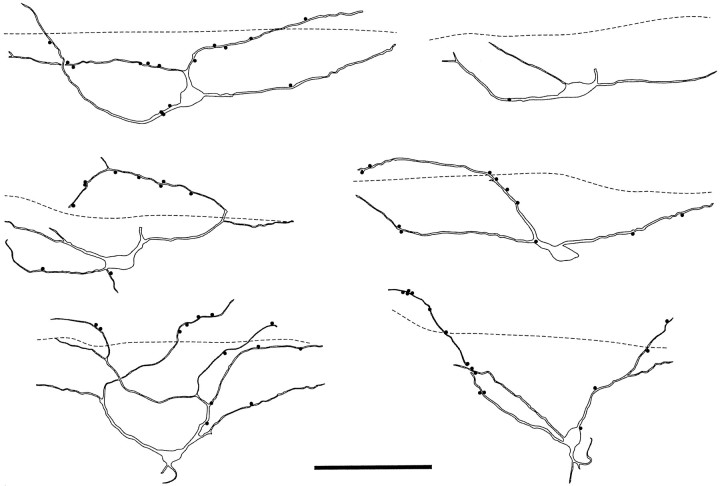
Fig. 4.
Drawings of six ChAT-immunoreactive neurons seen in parasagittal sections. In each case the dashed line represents the ventral limit of the plexus of substance P-containing axons, and filled circles indicate contacts from substance P-immunoreactive varicosities onto the cells. Scale bar, 100 μm.

