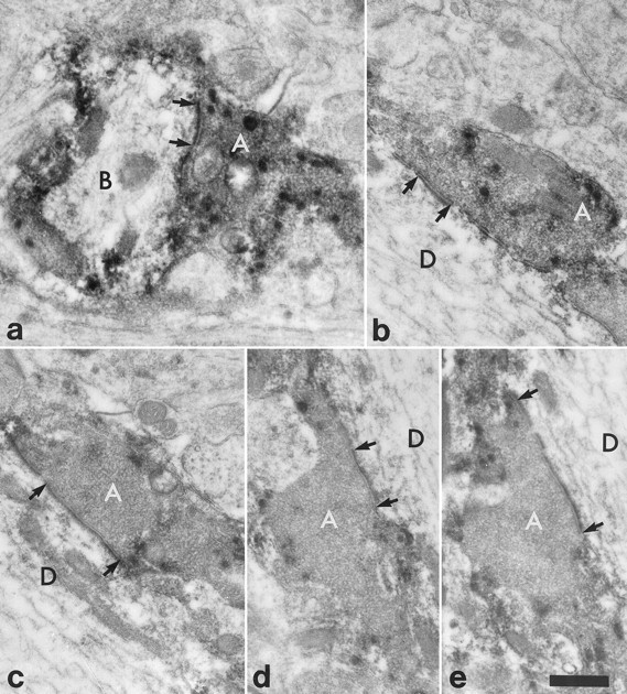
Fig. 7.

High-magnification electron micrographs to show synapses between the substance P-immunoreactive varicosities indicated with numbered arrows in Figure6b–d and the NK1 receptor-immunoreactive dendrite belonging to the cell illustrated in Figure 5. The micrographs are taken either from the ultrathin section illustrated in Figure6d (b, d) or else from nearby sections in the series (a, c, e).a, In a nearby section the substance P-immunoreactive axon (A; arrow 1 in Fig.6b–d) forms a synapse onto the small branch (B), which was given off from the main dendritic shaft. b–d, The substance P-immunoreactive axonal boutons (A) (numbered 2–4 in Fig.6b–d, respectively) form synapses onto the dendrite (D) of the NK1 receptor-immunoreactive neuron. e, The synapse shown in d is seen more clearly in a nearby ultrathin section. In each case an asymmetrical synaptic specialization is visible (between arrows). Scale bar, 0.5 μm.
