
Fig. 9.
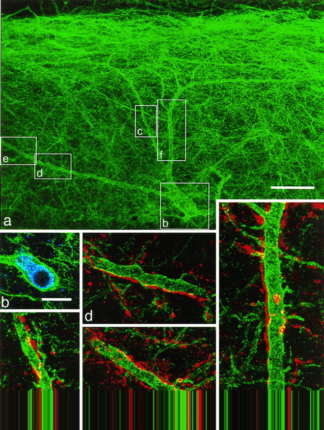
Substance P-immunoreactive contacts onto a spinothalamic neuron with the NK1 receptor. NK1 receptor immunoreactivity is shown in green(a–f), CTb immunoreactivity inblue (b), and substance P immunoreactivity in red (c–f).a, A low-magnification confocal image showing only the NK1 receptor immunoreactivity. The cell body is located 235 μm below the dorsal white matter, and dendrites extend up into the superficial dorsal horn. Boxes indicate the regions illustrated in b–f. b, NK1 receptor and CTb immunoreactivity in a single optical section through the cell body. c–f, Confocal images showing NK1 receptor and substance P immunoreactivity. The dendrites of this neuron receive numerous contacts from substance P-immunoreactive varicosities.a was obtained from nine optical sections 1.5 μm apart, c and e–f from four sections, andd from seven optical sections, each 0.5 μm apart. Scale bars: a, 50 μm; b, 20 μm;c–f, 10 μm.