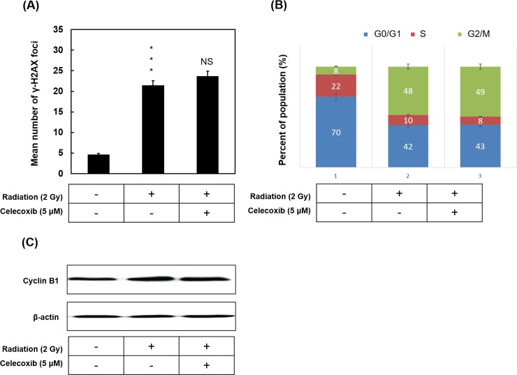
Fig 4. The effect of celecoxib and radiation on the formation of γ-H2AX foci and cell cycle distribution.
(A) A549 cells were treated with or without 5 μM Celecoxib followed by radiation of 2 Gy for 24 h. Cells were fixed and labeled with anti-γ-H2AX primary antibody and Alexa Fluor 555 conjugated secondary antibody. γ-H2AX foci were observed by fluorescence microscopy. Nuclei were counterstained with DAPI. The number of γ-H2AX foci per cell was counted at least 50 cells for each condition. The average number was expressed as the mean ± SD of three different experiments. ***P<0.001 indicated significant difference between control and radiation alone group. NS, no significance. (B) Cell cycle distribution in A549 cells with radiation treatment in combination with or without Celecoxib, and then analyzed by the flow cytometer. (C) Western blotting analysis of Cyclin B1 protein. β-Actin was used as loading control. Data are presented as the mean ± standard deviation, n = 3. ***P<0.001 vs. control. γ-H2AX, phosphorylated histone H2AX.

